Ultrasound
Yashoda Hospital offers state-of-the-art facilities to diagnose, evaluate, and manage a plethora of diseases, ailments, and malignancies. We have ultrasound machines that provide high-resolution images of the inside of the body. We possess high-end ultrasound machines that have excellent image quality and are equipped with modern applications like Harmonic Imaging, elastography, and 3D imaging.
What is Ultrasound?
Ultrasound imaging produces pictures of the body's internal structures by using a transducer or probe to generate sound waves. The procedure does not use ionizing radiation, provides a clear picture of soft tissues that are usually not presented well on X-ray images, and have known to have no harmful effects.
What are some common applications of the Ultrasound procedure?
Ultrasound examinations assist doctors in determining a variety of conditions and in assessing organ damage following symptoms such as:
- pain
- swelling
- infection
Ultrasound analyses various parts of body's internal organs, including but not limited to the:
- heart and blood vessels
- liver
- gallbladder
- spleen
- pancreas
- kidneys
- bladder
- ovaries, uterus, and fetus in pregnant patients
- eyes
- thyroid and parathyroid glands
- the scrotum (testicles)
- brain in infants
- hips in infants
- spine in infants
Ultrasound is also used to:
- oversee procedures such as needle biopsies where sample cells are taken from an abnormal area for laboratory testing.
- image the breasts and help in guiding breast cancer
- diagnose several heart conditions, including congestive heart failure and valve problems.
Doppler ultrasound images can support the physician to see and evaluate:
- blockages to blood flow
- narrowing of vessels
- tumors and congenital vascular malformations
- absent or reduced blood flow to various organs, such as the testes or ovary
- increased blood flow, which shows a sign of infection
How ultrasound scans work?
A small device called an ultrasound probe is used to create high-frequency sound waves inside the body. Images are created when these sound waves bounce off different parts of the body and generate "echoes" that are picked up by the probe. While the scan is being carried out, the images are displayed on the monitor.
How to Prepare for the Procedure?
Before the Procedure
- If you are going for an ultrasound in the pelvic area, you will be advised to drink water and not to go to the toilet until after the scan.
- If you are going for an ultrasound of the liver and gallbladder, you will be asked to avoid eating or drinking for several hours before the scan.
- You will be given a sedative to help you relax and inject a harmless substance called a contrast agent before the scan to generate clear images.
During the Procedure
- The radiologist or a sonographer will take 15 to 45 minutes to perform the tests.
- There are three main types of Ultrasounds scans, such as:
- External ultrasound scan: Here, the probe is moved over the skin
- Internal ultrasound scan: Here, the probe is inserted into the body
- Endoscopic ultrasound scan: Here, the probe is attached to a thin, long, and flexible tube that is passed deep inside the body.
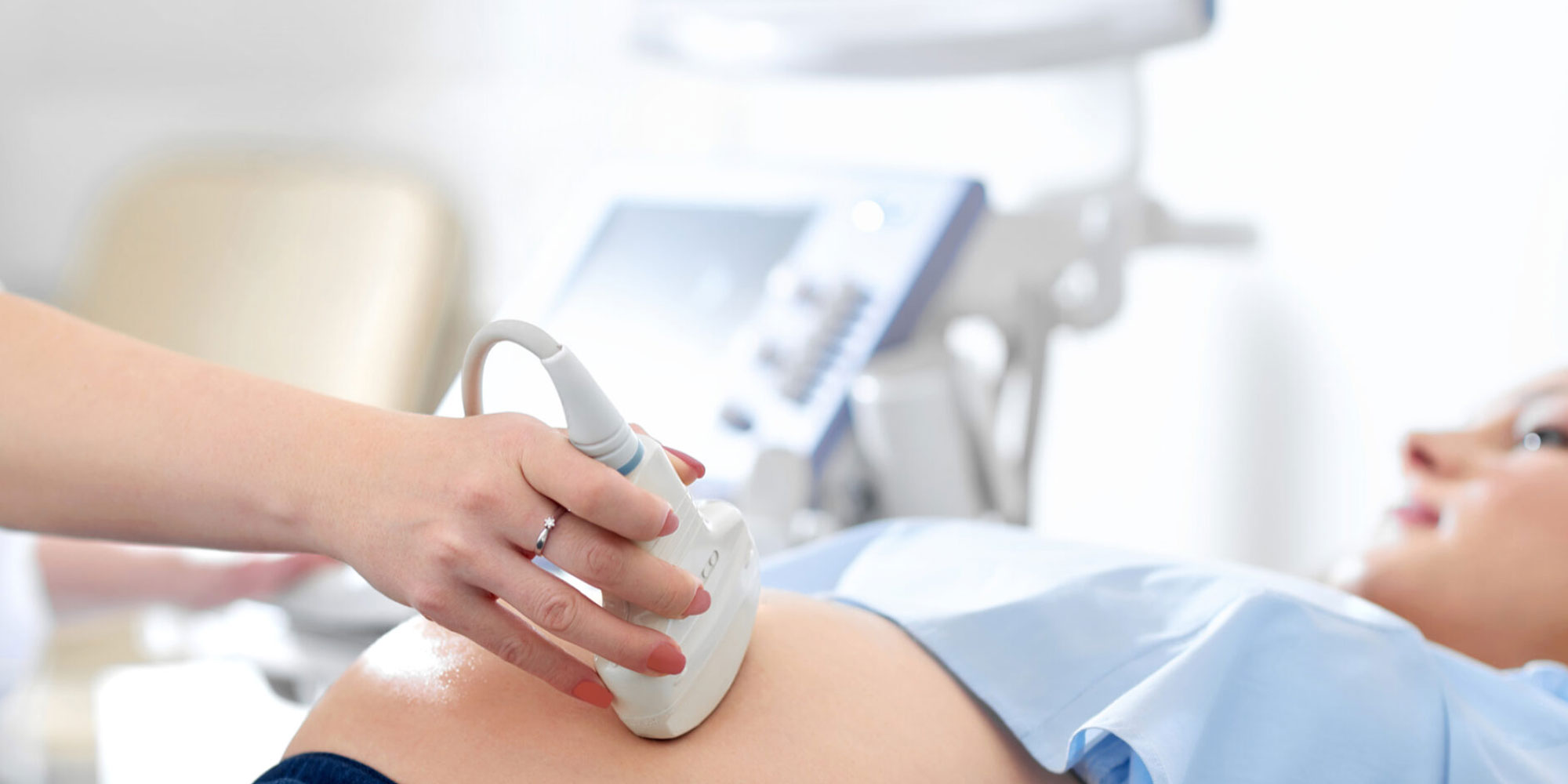
- You will be placed on the examination table. The radiologists or a trained sonographer will apply a water-based gel to the area which is being studied.
- The gel eliminates air pockets between the transducer and the skin by helping the transducer make secure contact with the body.
- The transducer is moved back and forth on the area until the intended images are captured.
- You will not experience any discomfort from the pressure as the transducer is pressed.
After the Procedure
- A radiologist, a doctor, trained to supervise and interpret radiology exams, will evaluate your images. The signed images will be shared with you and with your doctor.
- You may need to follow up on exams if there is a potential abnormality that needs further evaluation.
Risks
There are no known harmful effects on humans in standard diagnostic ultrasound.
Meet Our Doctors

