Digital Mammography at Yashoda Hospital
To detect breast cancer, mammography is the most common screening method. Our digital mammography and the Tomosynthesis system, located at Yashoda Radiology Department, can detect early breast cancer in patients than the previous procedures used in mammography technology. This procedure uses X-rays to help detect tumours or abnormal growths in the breasts. We offer latest advanced technologies for precision and accuracy to deliver quality health care.
What is Mammography?
Mammography is an X-ray picture of the breasts which utilizes a specialized medical imaging procedure. To identify early detection and diagnose breast medical conditions in women, a mammography exam, called a mammogram, is used by the doctors. Mammography is a non-invasive test that helps physicians diagnose and treat medical conditions.
The recent advances in mammography are- digital mammography and breast tomosynthesis.
What is Digital Mammography?
Digital mammography is also referred to as full-field digital mammography (FFDM), in which the x-ray film is not used, and instead, electronics are used to convert x-rays into mammographic pictures of the breast. This enables a lower radiation dose, and the images can also be transferred to a computer for review by the radiologist and for long term storage. The patient will have the same experience as those in the conventional film mammogram.
What is Breast Tomosynthesis?
Breast tomosynthesis, commonly called the three-dimensional (3-D) mammography, reconstruct three-dimensional images of the breasts by replacing X-rays with computers and using low-dose x-ray system.
Breast tomosynthesis supports the early detection and diagnosis of breast disease. Breast tomosynthesis has overcome some of the limitations of standard mammography.
Breast tomosynthesis has various advantages such as in:
- earlier detection of small breast cancers that may not be exhibited on a conventional mammogram
- fewer process of biopsies or additional tests
- considerable likelihood of identifying multiple breast tumors
- accurate images of abnormalities within dense breast tissue
- greater precision in pinpointing the shape, size, and location of breast abnormalities
How to Prepare for the Mammography Procedure?
Before the Procedure
- Before scheduling a mammogram, you should ask the doctor for advice.
- Try to avoid scheduling your mammogram for the week before your menstrual period because the breasts are usually tender during this time.
- Also, inform the radiologists if there is any possibility of you being pregnant.
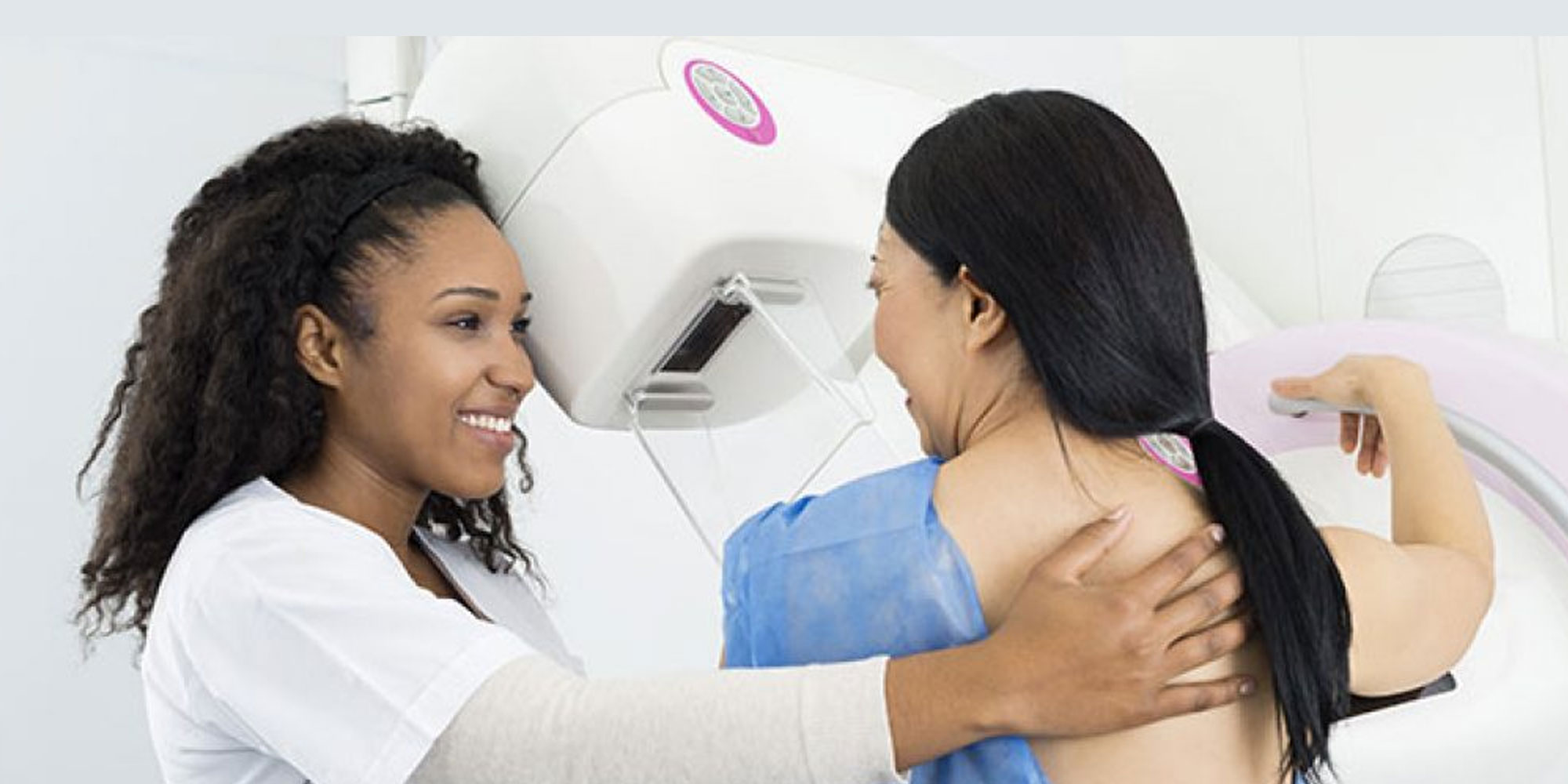
During the Procedure
- During mammography, an expert qualified radiologic technologist will place your breast in the mammography unit. A clear plastic paddle will compress your breasts, which are placed on a special platform.
- Breast compression is significant in order to:
- Even out the breast thickness to get visuals of all the tissue.
- Spread out the tissue so that any minor abnormalities that are hidden under overlying tissue could be exhibited.
- Get a thinner amount of breast tissue imaging by allowing the usage of a lower X-ray dose.
- Hold the breast still so that there are few blurred images caused by the motion.
- Reduce x-ray scatter to enhance the sharpness of the picture.
- You will have to change the position between images as the process will be repeated for the other breasts. You have to undergo compression for both digital mammography and tomosynthesis in order to minimize motion, which degrades the images.
- You will be asked to hold very still while the pictures are being taken to reduce the possibility of a blurred image.
- You may have to wait until after the examination is complete so that all the necessary images have been obtained.
- The entire examination process should take about 30 minutes.
After the Procedure
- Our trained physicians will supervise and interpret radiology examinations by analyzing the images.
- The signed reports will be shared with you or with your primary physician to discuss the results.
- You may have to undergo follow-up exams because of any potential abnormality needs, which may require evaluation with additional views or a special imaging technique.
- The doctor will ask you to go for further tests to see if the treatment is working.
Are there any risks from this procedure?
As with any type of X-ray, you are receiving exposure to some amount of radiation, but the risk is very low. Always inform your radiologists or technologists if you are pregnant.
Make an Appointment
We are eager to help you at every step of your medical journey from hospital and back to your home.
For Appointment
+91 - 9810922042
For Any Query
0120 – 4182000
Meet Our Doctors


