Fetal Anomaly Scan | Pregnancy Scan
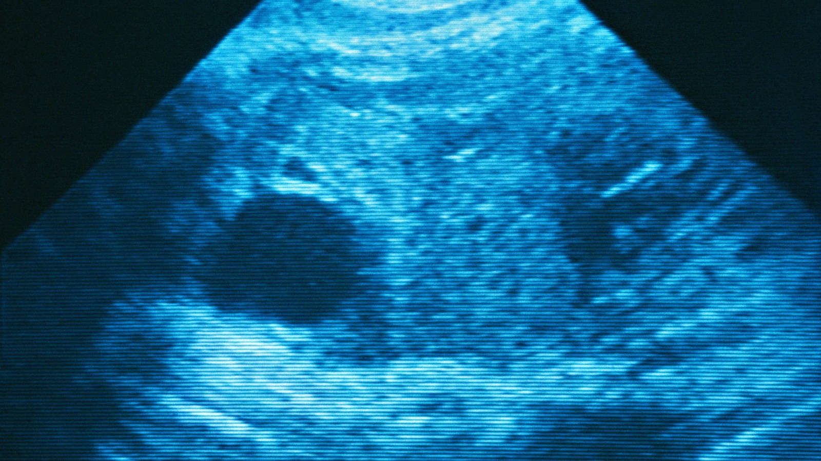
A fetal anomaly scan or mid-pregnancy scan is a detailed ultrasound scan performed between 18-21 weeks of pregnancy to take a close look at the uterus (womb) and the baby.
During the scan, the doctors examine each part of the fetal body including determination of the position of the placenta as well as assessing the amount of amniotic fluid, and measuring fetal growth.
What is the Purpose of a Fetal Scan?
The fetal anomaly scan is done to detect any physical abnormalities in the growing baby. Even though this scan cannot detect every abnormality in the fetus, it does provide a broad idea about the unborn baby’s bones, brain, heart, spinal cord, face, kidney and abdomen to the fetal medicine experts.
With this information, doctors can easily find out the following conditions:
- Anencephaly
- Diaphragmatic Hernia
- Gastroschisis
- Exomphalos
- Open Spina Bifida
- Bilateral Renal Agenesis
- Cleft Lips
- Edward’s Syndrome
- Patau’s Syndrome
- Serious Cardiac Abnormalities
Read Also: All About Women’s Doctor – Obstetrician-Gynecologist (OB-GYN)
Is a Fetal Scan Mandatory?
Before performing fetal scans, all pregnant women are informed about the procedure of the scan, its purpose and what it can and cannot detect. A pregnant woman’s consent is also necessary before performing the scan.
Types of Fetal Scans:
Modern technology can create both 2D and 3D fetal scans. These images can be produced either in black and white or in colour.
How is a Fetal Scan Performed?
During a fetal ultrasound, a specially trained healthcare professional, known as a sonographer, will follow these steps:
- Patient Positioning: The pregnant woman will be comfortably positioned on an examination table, typically lying on her back or side.
- Gel Application: A clear, hypoallergenic gel is then applied liberally to the patient’s abdomen (belly) so as to facilitate optimal sound wave transmission.
- Transducer Manipulation: The sonographer will utilize a handheld device called a transducer, gently gliding it across the gel-covered abdomen. The transducer emits and receives high-frequency sound waves that create images of the developing fetus.
- Image Acquisition: As the transducer moves, real-time two-dimensional (2D) black-and-white images of the fetus will be displayed on a monitor for examination.
- Hydration Optimization: In some cases, the sonographer may recommend increased fluid intake before the scan to ensure a full bladder, which can provide a clearer sonic window for visualizing the fetus.
- Pressure Application: Slight pressure may be applied on the transducer to achieve optimal image acquisition of certain fetal features.
Duration of the Fetal Scan:
The entire process of a fetal scan or mid-pregnancy scan takes about half an hour.
Areas of Focus in the Fetal Scan:
The sonographer will examine various body parts and organs of the baby and take measurements. Some of them are as follows:
- The sonographer may examine the shape and the size of the brain to check for brain problems.
- The sonographer may observe the face of the baby to check for problems like cleft lip.
- They may explore the spine to look for bone alignment.
- They may look at the abdominal wall to check whether it covers all the internal organs at the front or not.
- The sonographer may examine the heart of the baby to check for equal size of the atria and ventricles and also proper functioning of the valves.
- Kidneys are examined to check the proper functioning of the bladder.
- Hands and feet are examined to check the development of muscles, fingers and toes.
- The sonographer may also look at the position of the placenta.
- The sonographer may also check for the quantity of amniotic fluid for the baby’s free movement.
- The sonographer may also check the circumference of the head, abdomen and length of the femur by comparing and matching it with the standards of normal development.
Read Also: WHAT is Laparoscopic Myomectomy?
Which Abnormalities Can Be Seen in the Fetal Scan?
Here is a list of various abnormalities along with the likelihood of detection which the sonographer may observe in the fetal scan:
- Absence of the top of the head (anencephaly): 98 percent
- Cleft lip: 75 percent
- Abdominal defects
- Missing very short limbs: 60 percent
- Spinal defects: 90 percent
- Major kidney problems: 84 percent
- Chromosomal abnormalities: 95 percent
- Major heart problems: 50 percent
Results of the Scan:
In most of the fetal scans, no abnormalities are picked up and most of the babies develop normally. The sonographer may take an opinion from an experienced colleague or a specialist when a problem is detected.
Though most of the abnormalities are traceable through fetal scans, still there are chances that the baby may be born with an anomaly that had gone previously undetected.
Read Also: Types of Diabetes: Meaning, Symptoms, Causes, Treatment & Prevention
What Happens in Case a Problem is Detected?
Doctors at Yashoda Hospital & Research Centre, Nehru Nagar, Ghaziabad, may order various other tests or scans to understand the problem better. Once the anomaly is confirmed, further course of action depends on its seriousness. The less serious anomalies may get better with time.
In case of serious problems, the doctors and specialists may provide support and information
to the family about all the possible courses of action including termination of pregnancy. The family can make a choice that is always respected.
Read Also: How to Control Diabetes In Pregnancy
Conclusion:
Early detection of an anomaly before birth can help in planning post-birth treatment. This may be beneficial in cases wherein surgery is required immediately after birth as it increases the chances of survival of the baby.
Pregnant women can consult doctors at Yashoda Hospital & Research Centre, Nehru Nagar, Ghaziabad, as they will make sure that you and your baby are healthy and active via regular checkups throughout the pregnancy.
Meet Obstetricians & Gynecologists who will assist you with all tests you need to rule out the abnormalities.
Dr. Ranjana Mishra is currently a Senior Consultant in Medical Genetics at Yashoda Hospital & Research Centre, Nehru Nagar, Ghaziabad. Dr. Mishra brings over 17 years of expertise in obstetrics and gynecology across various sectors, along with a unique specialization in medical genetics. She combines precision and care to redefine women’s health through early screening for genetic diseases.